
Zoology‚ through hands-on exploration‚ unveils life’s diversity; this laboratory manual‚ alongside Zoology by Miller‚ guides students through practical investigations and detailed observations.
The Scope of Zoology
Zoology encompasses the vast and intricate study of the animal kingdom‚ extending from microscopic cellular structures to complex ecosystems and evolutionary histories. This discipline delves into animal anatomy‚ physiology‚ behavior‚ ecology‚ distribution‚ and classification. Laboratory studies are pivotal‚ offering direct engagement with animal diversity – from the simplest sponges (Porifera) to sophisticated mammals.
Practical exploration‚ as guided by resources like Stephen A. Miller’s Zoology and accompanying laboratory manuals‚ allows students to dissect‚ observe‚ and analyze specimens. This hands-on approach fosters a deeper understanding of animal form and function. The scope extends to invertebrate zoology‚ examining creatures like jellyfish (Cnidaria)‚ flatworms (Platyhelminthes)‚ and segmented worms (Annelida)‚ alongside vertebrate studies involving fish‚ amphibians‚ reptiles‚ birds‚ and mammals. Ultimately‚ zoology seeks to unravel the fundamental principles governing animal life.
Importance of Laboratory Work in Zoology
Laboratory work is fundamental to a comprehensive zoology education‚ bridging theoretical knowledge with practical application. Direct observation and dissection‚ as facilitated by a laboratory manual alongside texts like Miller’s Zoology‚ cultivate critical thinking and analytical skills. Students move beyond textbook descriptions‚ engaging directly with animal anatomy and physiology.
These experiences refine observational abilities‚ essential for identifying structures and understanding functional relationships. Furthermore‚ lab work reinforces concepts of classification‚ adaptation‚ and evolution. Examining specimens – from invertebrates like sponges and worms to vertebrates like fish and mammals – provides tangible evidence supporting evolutionary theory. The ability to meticulously record observations‚ draw biological structures‚ and analyze data‚ as emphasized in introductory courses (Biology 1413)‚ are invaluable skills honed through laboratory practice‚ preparing students for advanced zoological studies and research.
Laboratory Safety Protocols
Prioritize safety! Familiarize yourself with equipment and supplies‚ adhering to guidelines detailed in supplemental materials like Ziser’s 2015.12 laboratory safety document.

General Safety Guidelines
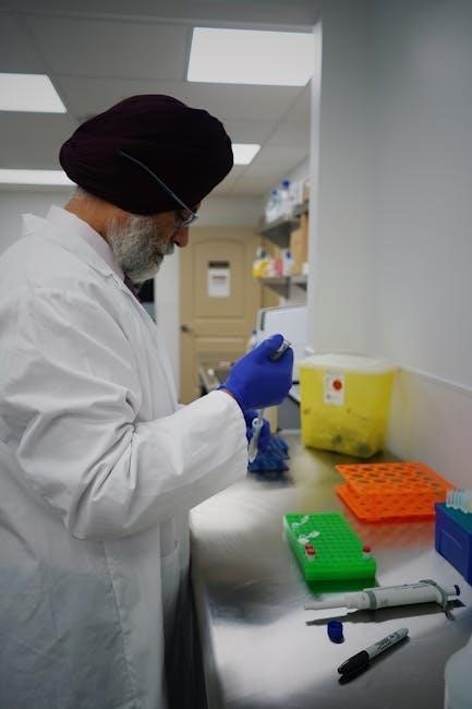
Prioritizing safety is paramount within the zoological laboratory. Always wear appropriate personal protective equipment (PPE)‚ including lab coats‚ gloves‚ and eye protection‚ to minimize exposure to potential hazards. Maintain a clean and organized workspace‚ promptly addressing any spills or broken glassware.
Be mindful of your surroundings and the activities of others‚ fostering a collaborative yet cautious environment. Never eat‚ drink‚ or apply cosmetics in the lab‚ preventing accidental ingestion of potentially harmful substances. Thoroughly wash your hands with soap and water after handling specimens or completing experiments.
Report any accidents‚ injuries‚ or unsafe conditions to the instructor immediately. Understand the location and proper use of safety equipment‚ such as fire extinguishers and eyewash stations. Adherence to these guidelines ensures a productive and secure learning experience for everyone involved in zoological studies.
Handling of Specimens
Proper specimen handling is crucial for both preservation and safety. Always treat biological materials with respect‚ recognizing their origin and potential hazards. Utilize appropriate tools – forceps‚ dissecting scissors‚ probes – to minimize direct contact. When dissecting‚ follow established protocols carefully‚ making precise incisions and observing anatomical structures systematically.
Preserved specimens often contain formaldehyde or other fixatives; handle them with gloves and in a well-ventilated area. Avoid damaging delicate tissues during manipulation. Document observations meticulously through drawings or photographs‚ noting key features and characteristics.
Return specimens to their designated containers after use‚ ensuring proper labeling and storage. Never remove specimens from the laboratory without authorization. Responsible handling safeguards both the integrity of the specimens and the well-being of researchers.
Disposal of Biological Waste
Safe and responsible disposal of biological waste is paramount in a zoological laboratory. Follow established protocols meticulously to prevent contamination and ensure environmental protection. Sharps – slides‚ cover slips‚ scalpels‚ and needles – must be discarded in designated puncture-resistant containers‚ never in general trash.
Dissected specimens‚ tissues‚ and other biological materials should be placed in biohazard bags‚ clearly labeled‚ and autoclaved or chemically disinfected before disposal. Liquid waste containing biological agents requires specific treatment‚ often involving filtration or sterilization.
Always adhere to institutional guidelines and local regulations regarding biohazard waste management. Never pour biological waste down the drain. Proper disposal minimizes risks to personnel‚ the environment‚ and the broader community‚ upholding ethical laboratory practices.

Microscopy Techniques
Microscopy is central to zoological study‚ enabling visualization of cellular structures and tissues; mastering techniques like slide preparation is crucial for accurate observation.
Types of Microscopes Used in Zoology
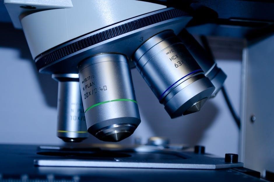
Zoological laboratories commonly employ several microscope types‚ each suited for specific observational needs. The compound microscope‚ a foundational tool‚ utilizes multiple lenses to achieve high magnification‚ ideal for examining cells and tissues. Stereo microscopes‚ also known as dissecting microscopes‚ provide a three-dimensional view‚ beneficial for observing surface features and performing dissections.

Beyond these‚ phase-contrast microscopes enhance the visibility of transparent specimens without staining‚ revealing intricate cellular details. Electron microscopes‚ including Transmission Electron Microscopes (TEM) and Scanning Electron Microscopes (SEM)‚ offer significantly higher resolution‚ allowing visualization of ultrastructural components like organelles and viruses. TEM examines internal structures‚ while SEM provides detailed surface imaging.
Fluorescence microscopy utilizes fluorescent dyes to highlight specific cellular components‚ aiding in specialized studies. The choice of microscope depends on the specimen‚ desired magnification‚ and the specific features being investigated‚ making a diverse range of options essential for comprehensive zoological research.
Preparing Slides for Microscopic Observation
Effective microscopic observation hinges on proper slide preparation. Wet mounts are quick and simple‚ ideal for temporary observations of living specimens; a drop of liquid containing the sample is placed on a slide and covered with a coverslip. For permanent slides‚ fixed specimens undergo preservation and embedding in paraffin or resin.
Sectioning‚ using a microtome‚ creates thin slices for optimal light transmission. These sections are then mounted on slides and stained with dyes like hematoxylin and eosin to enhance contrast and reveal cellular structures. Staining techniques vary depending on the tissue type and features of interest.
Proper mounting ensures the specimen remains flat and protected. Careful handling of coverslips prevents air bubbles and ensures clear visualization. Accurate labeling is crucial for identification and record-keeping. Mastering these techniques is fundamental for successful zoological investigations.
Magnification and Resolution
Magnification‚ the degree to which an image appears larger‚ is a key function of microscopes‚ achieved through the objective and ocular lenses. However‚ magnification alone isn’t sufficient; resolution – the ability to distinguish between two closely spaced points – is equally vital for clear‚ detailed images.

Higher magnification doesn’t always equate to better resolution. Resolution is limited by the wavelength of light used and the numerical aperture of the objective lens. Oil immersion lenses increase resolution by reducing light refraction. Understanding these principles is crucial for interpreting microscopic observations.
Proper illumination and focusing are essential to maximize both magnification and resolution. Careful adjustment of the condenser and light source optimizes image clarity. Recognizing the interplay between these factors allows for accurate and insightful zoological studies.

Invertebrate Zoology: A Practical Approach
Invertebrate studies‚ guided by manuals like Dr. Drews’‚ involve detailed examinations of diverse phyla – Porifera‚ Cnidaria‚ Platyhelminthes‚ Nematoda‚ and Annelida – revealing their unique adaptations.
Study of Porifera (Sponges)
Porifera‚ commonly known as sponges‚ represent the simplest multicellular animals‚ exhibiting a unique body plan lacking true tissues or organs. Laboratory investigations focus on observing their structural characteristics‚ including the presence of spicules – skeletal elements composed of calcium carbonate or silica – which aid in identification. Students will examine sponge morphology‚ noting the ostia (pores) through which water enters and the osculum‚ the large opening for water expulsion.
Dissection‚ though limited due to their simple structure‚ allows for visualization of the internal canal system responsible for nutrient acquisition and waste removal. Microscopic observation reveals the choanocytes‚ flagellated cells that generate water currents and capture food particles. Understanding sponge anatomy highlights their evolutionary significance as early diverging metazoans‚ demonstrating fundamental principles of cellular organization and specialization. Careful observation and detailed drawings are crucial for documenting these features.
Observation of Cnidaria (Jellyfish‚ Corals)
Cnidaria‚ encompassing jellyfish‚ corals‚ and sea anemones‚ exhibit radial symmetry and possess specialized stinging cells called cnidocytes. Laboratory studies involve observing both polyp and medusa forms‚ noting their distinct morphologies and life cycle stages. Prepared slides of Hydra‚ a freshwater polyp‚ allow for detailed examination of its tentacles‚ gastrovascular cavity‚ and budding reproductive structures.
Jellyfish dissection reveals the gelatinous mesoglea‚ the bell-shaped body‚ and the oral arms equipped with cnidocytes. Microscopic observation of cnidocytes demonstrates the mechanism of nematocyst discharge – a rapid expulsion of a stinging thread used for prey capture and defense. Coral skeletons‚ representing colonial cnidarians‚ showcase diverse growth forms and calcium carbonate structures. Understanding cnidarian biology illuminates their ecological roles and evolutionary adaptations within marine ecosystems.
Examination of Platyhelminthes (Flatworms)
Platyhelminthes‚ or flatworms‚ represent the simplest bilaterally symmetrical animals. Laboratory investigations focus on identifying key characteristics like dorsoventral flattening‚ incomplete digestive systems‚ and protonephridia for osmoregulation. Planaria‚ a free-living flatworm‚ serves as an excellent model for observing regeneration capabilities; fragmentation and subsequent regrowth demonstrate remarkable plasticity.
Microscopic examination of Planaria reveals the cellular structure of the body‚ including the gut‚ pharynx‚ and flame cells. Parasitic flatworms‚ such as tapeworms (Taenia) and flukes‚ are studied using prepared slides‚ highlighting their specialized adaptations for host exploitation – scolexes with hooks and suckers‚ and complex reproductive systems. Observing these features underscores the ecological and medical significance of Platyhelminthes.
Analysis of Nematoda (Roundworms)
Nematoda‚ commonly known as roundworms‚ are pseudocoelomates exhibiting cylindrical‚ unsegmented bodies. Laboratory studies emphasize their adaptation to diverse environments‚ from soil to parasitic lifestyles within plants and animals. Ascaris lumbricoides‚ a prevalent intestinal parasite‚ is frequently examined to illustrate their reproductive strategies – separate sexes and prolific egg production.
Dissection of preserved Ascaris specimens reveals a complete digestive system with a mouth‚ pharynx‚ intestine‚ and anus. Microscopic observation focuses on identifying key features like the cuticle‚ pseudocoelom‚ and longitudinal muscles. Furthermore‚ parasitic nematodes like hookworms and pinworms are studied via prepared slides‚ showcasing their specialized structures for attachment and nutrient absorption within their hosts. Understanding Nematoda’s morphology and life cycles is crucial for comprehending their ecological roles and impacts on human and animal health.

Exploring Annelida (Segmented Worms)
Annelida‚ or segmented worms‚ demonstrate a significant evolutionary advancement with their metameric bodies. Laboratory investigations typically center on the earthworm‚ Lumbricus terrestris‚ to illustrate annelid anatomy and physiology. Dissection reveals a well-developed coelom‚ segmented internally and externally‚ providing hydrostatic support and enabling efficient locomotion.
Students examine the digestive system‚ noting the specialized structures like the crop‚ gizzard‚ and intestine. The circulatory system‚ featuring dorsal and ventral blood vessels‚ and the nervous system‚ with a cerebral ganglion and ventral nerve cord‚ are also key areas of study. Observing the nephridia‚ excretory organs in each segment‚ highlights their role in osmoregulation. Prepared slides of marine annelids‚ like sandworms‚ showcase parapodia and setae‚ adaptations for aquatic life. Analyzing Annelida provides insight into the evolution of segmentation and complex organ systems.

Vertebrate Zoology: Dissection and Observation
Vertebrate studies involve detailed dissections‚ revealing anatomical structures and physiological functions; comparative analysis illuminates evolutionary adaptations across fish‚ amphibians‚ reptiles‚ birds‚ and mammals.
Fish Dissection: Anatomy and Physiology
Fish dissection provides a foundational understanding of vertebrate anatomy‚ initiating students into the complexities of organ systems. Careful observation reveals external features – scales‚ fins‚ lateral lines – adapted for aquatic life. Internal dissection exposes the streamlined body plan‚ including the digestive tract‚ circulatory system with a two-chambered heart‚ and respiratory system utilizing gills for efficient oxygen extraction.
Students will identify key structures like the swim bladder‚ crucial for buoyancy control‚ and examine the nervous system‚ noting the relatively small brain compared to higher vertebrates. The reproductive system‚ varying between species‚ offers insights into fish life cycles. Analyzing muscle arrangement demonstrates adaptations for swimming. This practical experience correlates directly with physiological functions‚ illustrating how anatomy dictates performance in an aquatic environment‚ and builds a strong base for comparative vertebrate studies.
Amphibian Study: External and Internal Features
Amphibian study bridges aquatic and terrestrial adaptations‚ showcasing a fascinating evolutionary transition; External examination reveals moist‚ permeable skin facilitating cutaneous respiration‚ alongside limbs adapted for both swimming and walking. Observing the head highlights features like prominent eyes and a tympanic membrane for detecting sound. Internal dissection unveils a three-chambered heart‚ less efficient than those of reptiles or mammals‚ but sufficient for their metabolic needs.
Students will trace the digestive system‚ noting adaptations for a carnivorous diet‚ and examine the respiratory system‚ including lungs and the aforementioned skin. The urogenital system demonstrates adaptations for both aquatic reproduction and waste excretion. Analyzing the nervous system reveals a more developed brain compared to fish. This practical exploration illuminates the physiological challenges and evolutionary solutions associated with a semi-aquatic lifestyle‚ providing valuable comparative data.
Reptile Examination: Adaptations and Characteristics
Reptile examination focuses on adaptations for fully terrestrial life‚ contrasting sharply with amphibians. Key external features include dry‚ scaly skin minimizing water loss‚ and lungs as the primary respiratory organ. Observe limb structure‚ varying significantly based on locomotor style – sprawling in lizards‚ more upright in crocodiles. Internal dissection reveals a more efficient three-chambered (or four-chambered in crocodilians) heart‚ supporting higher metabolic rates.
Trace the digestive system‚ noting adaptations for diverse diets‚ and examine the excretory system‚ highlighting the production of uric acid to conserve water. The amniotic egg‚ a defining reptilian characteristic‚ should be conceptually understood. Analyzing the nervous system reveals further brain development. This practical study illuminates the physiological and anatomical features enabling reptiles to thrive in diverse terrestrial environments‚ offering crucial comparative insights.
Bird Anatomy: Flight and Feather Structure
Bird anatomy uniquely adapts to flight‚ demanding lightweight yet strong structures. Dissection reveals pneumatic bones‚ hollow and reinforced‚ reducing weight. Observe the keeled sternum‚ providing a large surface for flight muscle attachment. Wing structure is paramount; examine the different feather types – contour‚ flight‚ and down – noting their roles in aerodynamics and insulation.
Trace the highly efficient respiratory system‚ including air sacs extending throughout the body‚ facilitating unidirectional airflow. The avian heart‚ with its four chambers‚ supports high metabolic demands. Investigate the digestive system‚ adapted for rapid processing of energy-rich food. Analyze the skeletal adaptations‚ particularly the fused bones‚ enhancing flight stability. Understanding these features illuminates the evolutionary success of birds.
Mammalian Dissection: Organ Systems
Mammalian dissection provides a comprehensive view of complex organ systems. Begin with external features‚ noting adaptations related to locomotion and feeding. Systematically dissect to reveal the muscular‚ skeletal‚ and integumentary systems‚ observing their interrelationships. The digestive system demonstrates specialized regions for processing food; trace its path from mouth to anus.
Examine the cardiovascular system‚ identifying the four-chambered heart and major blood vessels. Investigate the respiratory system‚ focusing on the lungs and diaphragm. The urogenital system reveals adaptations for waste removal and reproduction. Finally‚ explore the nervous system‚ identifying the brain‚ spinal cord‚ and peripheral nerves. Careful observation and accurate identification are crucial for understanding mammalian physiology.
Data Recording and Analysis
Laboratory notebooks are essential for detailed observations‚ sketches of biological structures‚ and statistical analysis of zoological data‚ ensuring accurate scientific reporting.
Maintaining a Laboratory Notebook
A meticulously maintained laboratory notebook serves as a primary record of your zoological investigations‚ functioning as a detailed chronicle of experiments‚ observations‚ and analyses. Each entry should begin with the date and a clear‚ concise title describing the experiment or observation. Record all procedures with sufficient detail to allow replication by another researcher – include materials used‚ specific methodologies followed‚ and any deviations from established protocols.
Observations should be recorded immediately and objectively‚ noting both expected and unexpected results. Utilize descriptive language‚ diagrams‚ and‚ where appropriate‚ quantitative data. Avoid interpretations or conclusions within the raw data section; these belong in the analysis portion. Ensure all entries are written in permanent ink and are legible. Number pages consecutively and avoid leaving blank spaces – cross out any errors with a single line‚ rather than erasing‚ and initial the correction. A well-maintained notebook is not merely a record‚ but a testament to the scientific process itself.
Drawing Biological Structures
Biological illustration is a crucial skill in zoology‚ enhancing observational abilities and facilitating accurate record-keeping. Drawings should be executed with a sharp pencil on white‚ unlined paper‚ prioritizing clarity and precision over artistic flair. Begin with light‚ preliminary lines to establish proportions and overall shape‚ then gradually refine details.
Label all significant structures directly on the drawing using leader lines – avoid crowding labels. Stippling or hatching techniques are preferred for shading‚ conveying texture and depth without obscuring underlying structures. Maintain proper proportions and relative sizes; avoid overly detailed renderings that obscure essential features. Include a descriptive title and magnification level. Practice regularly to improve your skills; accurate drawings are invaluable for understanding and communicating complex biological forms‚ supplementing photographic documentation.
Statistical Analysis of Zoological Data
Statistical methods are essential for interpreting zoological data‚ moving beyond descriptive observations to draw meaningful conclusions. Common techniques include calculating measures of central tendency (mean‚ median‚ mode) and dispersion (standard deviation‚ variance) to summarize data sets.
T-tests compare means between two groups‚ while ANOVA analyzes variances among multiple groups. Chi-square tests assess relationships between categorical variables. Understanding p-values is crucial; a p-value less than 0.05 typically indicates statistical significance‚ suggesting the observed results are unlikely due to chance. Appropriate statistical tests depend on the data type and research question. Software packages like R or SPSS can facilitate these analyses. Careful consideration of sample size and potential biases is vital for valid interpretations‚ ensuring robust conclusions in zoological research.